Clarifying the Role of Vitamin E
“Scientists have struggled for decades to determine the role that Vitamin E plays in human health.” Scientists have debated the…

Researchers at the University of Toronto and University of Regina are using neutron imaging to reveal the invisible structures of soft tissues inside fossils.
The most valuable information in a fossil is often the hardest to access.
Buried within stone are the structures that reveal how extinct animals lived—cartilage, blood vessels, and other soft tissues that rarely survive fossilization in a form that can be studied. For decades, paleontologists assumed that such materials were completely lost over time.
Only in the past 20 years has it become clear that traces of soft tissue can persist in ancient fossils, and the scientific world is still discovering the wealth of information to be learned about extinct creatures from them.
Yet a fundamental challenge remains: these structures are hidden deep within fossilized bone, and conventional techniques either cannot reveal them or risk damaging the specimen.
Because of these challenges, the interior of most fossils has not been systematically examined for soft tissues using non-destructive methods. As a result, the world’s collections of fossils from millions of individual organisms, including tens of thousands of dinosaurs, remain to be screened for presence of soft issues—a treasure trove for discovery.
With the growing abundance of well-preserved fossil specimens held in Canadian collections, the ability to non-destructively screen these materials represents a significant scientific and national cultural opportunity.
Neutron and synchrotron x-ray imaging offer means to look inside fossils without cutting into them. Neutron imaging can penetrate deeper into dense fossil material than x‑rays, which is especially valuable for dinosaur bones, some of which are quite large. These non-destructive tools both require large-scale facilities.
Two independent Canadian-led efforts illustrate how neutron imaging is becoming a transformative tool for examination of fossils: one led by the University of Regina and another by the University of Toronto.
Jerit Mitchell, a PhD researcher at the University of Regina, and his research supervisors—Prof. Mauricio Barbi and Prof. Marcella Berg— are searching for preserved soft tissues in fossilized bones, and in the process are building a pipeline of paleontological discovery based on neutron imaging and synchrotron x-ray methods to study fossilized bone.

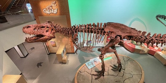
His work on Scotty, a Tyrannosaurus rex on display at the Royal Saskatchewan Museum, showed how synchrotron x-ray imaging can reveal vascular structures associated with bone healing, providing insight into dinosaur physiology.
Now, he has taken neutron images to detect signatures of soft tissue preservation within fossilized bone. He is looking for insights into these extinct animals’ blood vessels and injury-healing abilities, as well as the phenomenon of ancient soft tissue preservation in general.

“Neutron imaging gives us a way to look for soft tissue signals that are simply invisible to other techniques,” as Mitchell explains. “In addition to being more penetrating than x-rays, neutrons are much more sensitive to the hydrogen-rich materials that make up soft tissues of all living things. If there is any soft tissue remaining deep inside large dinosaur fossils, we are likely to see it with neutrons.”
He is pioneering the combination of synchrotron x-ray and neutron imaging to search for these tissues and glean insights. Such explorations mark a critical transition from inference to observation. Where earlier work without the soft tissues relied on indirect evidence—morphology and chemical signatures—these non-destructive imaging techniques offer the possibility of directly visualizing contrasts linked to preserved biological material.
A complementary effort at the University of Toronto focuses on other reptiles and vertebrates. Professor Robert Reisz is not new to pushing the boundaries of what fossils can reveal. He is a leading authority on vertebrate paleontology, particularly the evolution of reptiles and early amniotes. Reisz is a Fellow of the Royal Society of Canada in recognition of his substantial, sustained contributions to science.

In recent years, Reisz has increasingly turned to neutron facilities to study fossils that resist conventional approaches. His work—often in collaboration with the Australian Centre for Neutron Scattering (ACNS)—has applied neutron tomography to multiple fossil systems, particularly Permian vertebrates preserved in dense or chemically complex matrices, where x‑ray imaging struggles and destructive preparation is not an option. Many of these fossils, found by Reisz and donated to Royal Ontario Museum, have formed the basis of a vibrant research program using neutron imaging at ACNS.

In one of the most striking recent examples, this approach has made it possible to examine a fossil that would otherwise remain inaccessible. The specimen—areptile (Captorhinus) radiometrically dated at nearly 300 million years—is extraordinary for its preservation. Unlike typical fossils, it retains not only bone but also three-dimensional skin, cartilage, and remnants of original protein structures, offering a rare snapshot of soft tissue anatomy of extinct reptiles.
The challenge was immediate: these preserved features are precisely the ones most likely to be destroyed by traditional fossil preparation.
At ACNS, neutron imaging allowed the research team to image the fossil, reconstructing internal structures in three dimensions without physically altering the specimen. The resulting data revealed details of the ribcage, cartilage, and skeletal articulation, providing new insight into how these terrestrial vertebrates breathed.
The importance of neutron imaging in this case is not incremental—it is enabling.
“Only neutron imaging has the unique ability to visualise these extremely delicate soft tissues preserved within the rock,” says Prof. Robert Reisz.
“Neutron imaging is moving fossil studies beyond bone and into preserved biology, revealing anatomical features that would otherwise remain hidden—or be lost entirely—allowing questions about physiology and function to be addressed directly.”
The work of Reisz and Mitchell illustrates a shift in paleontology. Neutron imaging has already been applied across multiple fossil systems to reveal structures that could not be accessed any other way.
Canadian researchers are pushing neutron methods into new territory—probing dinosaur and other ancient fossils for traces of soft tissue that have remained hidden until now.
These methods are not simply improving resolution—they are changing the scope of what can be studied.
But they also reveal a structural reality. The ability to carry out this work depends on access to specialized facilities—facilities that are scarce, in high demand, and often located outside Canada.
The result is a research environment in which scientific opportunity is closely tied to infrastructure access.
As a result, Canadian researchers have very limited access to the small number of large-scale facilities globally. While Reisz relies on an Australian facility, Mitchell has managed to get sporadic access to some European and American facilities.
Some of his images of dinosaur fossils were taken through Canada’s limited-time partnership with the ISIS Neutron and Muon Source in the United Kingdom. Although Canada is not a member of the Institut Laue-Langevin, a multi-national neutron source in France, because of the novelty and importance of this research, he was able to obtain temporary access to demonstrate its potential. He also had recent access to beamlines at Oak Ridge National Lab in the United States.
“We’ve been fortunate in getting beam time so far,” as Mitchell explains, “But with so many fossils to screen for soft tissues, regular and consistent access is needed. Also, going abroad adds a layer of complexity—especially when you are transporting rare and fragile specimens—there’s a higher risk of damage in transit. And you expend a lot more time and expense for travel for every experiment.”
While Reisz has successfully developed an on-going collaboration with ACNS, taking fossils as far as Australia results in inevitable losses. He has started to hand-carry specimens when he travels there after a shipper lost some of his valuable materials.
At present, the ability of Canadian researchers to systematically explore one of the world’s most valuable fossil collections is effectively limited.
A Canadian neutron imaging facility could alleviate many of these problems.
As neutron imaging begins to open new windows into the fossil record—revealing physiology, preservation pathways, and biological detail once thought lost—the question is no longer whether neutron imaging can transform paleontology.
It is whether Canadian researchers will have reliable access to the neutron beams required to do it.